Seeing is believing: The cutting edge of watching single molecules inside human cells
The cell is a complex network of interacting components, or molecules, each of them with its own characteristics and all of them together functioning as a living system. Each of the molecular processes and interactions in the cell bears the risk of becoming dysfunctional, resulting in disease. Biomedical research into processes that power the cell lays the foundation for major therapeutic breakthroughs. However, the minuscule length scales and high speeds at which these intracellular processes take place make it very challenging to observe them directly within a single cell.
Nils Walter’s team at the University of Michigan, Chemistry Department and Center for RNA Biomedicine, has reviewed the latest research on using high-resolution, single molecule fluorescence microscopy tools to study the interactions between molecules in live human cells in real time. The review covers research reported in 85 publications over the last 5 years, aiming to consolidate the developments and disseminate the techniques that can follow multiple molecules at once (“multiplexing”). These techniques are relatively easy to implement, and are becoming increasingly available and affordable.
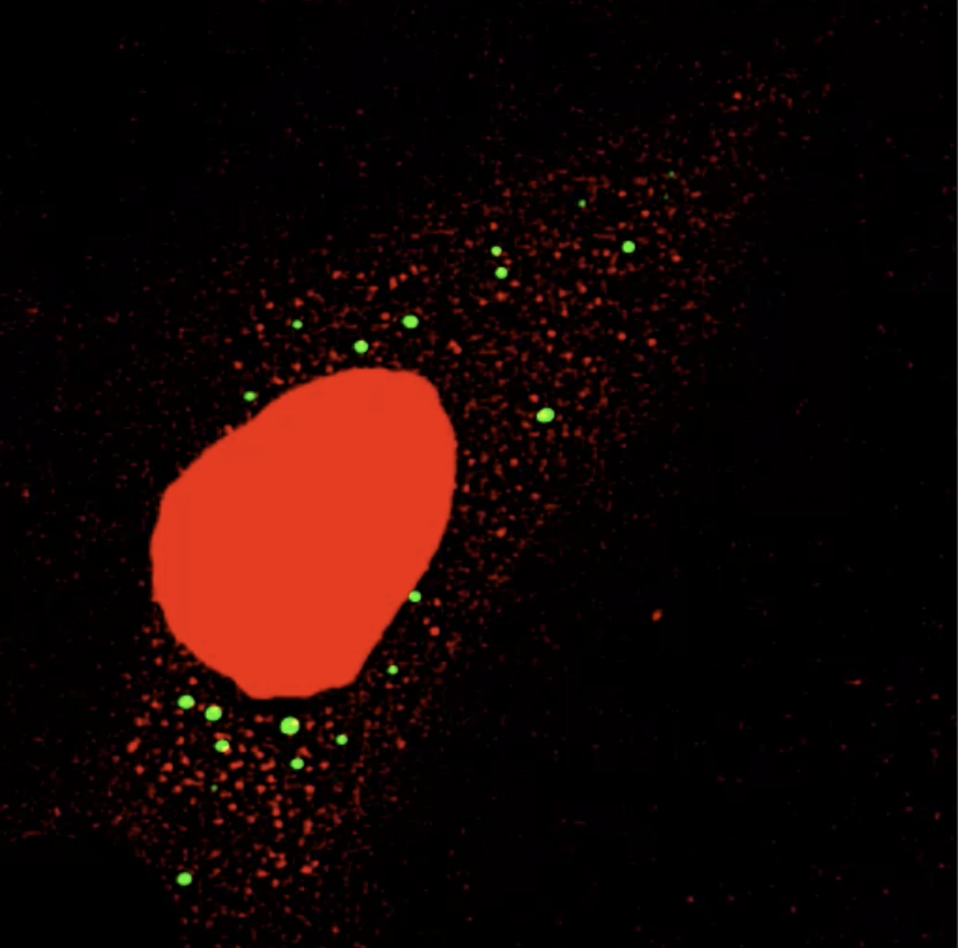
Red RNA molecules are docking onto green processing bodies containing RNA degrading enzymes.
Two major technological advances have been transforming the field of single molecule, single cell studies. Advances in fluorescence microscopy and clever engineering now offer very high-sensitivity and high-resolution capabilities, allowing researchers to see and record intracellular interactions using a growing toolbox of fluorescent tags to track cellular components. At the same time, the field of RNA biology has made great strides in understanding the link between aberrant gene regulation and disease.
Multiplexed techniques, able to follow multiple molecular species, are being progressively applied to intracellular contexts, becoming more biologically relevant as they get closer to disease physiology.
“Multiplexing is the opportunity to look at cell component interactions,” explains Saffron Little, one of the five co-authors of this review. “And the advancement of these technologies has allowed us to view these interactions in real time.”
“Most labs could now easily uptake multiplexed technology” says Andreas Schmidt, the lead author of this article. “We’d like to see the adoption of this technology speed up, because it can lead to major therapeutic discoveries.”
Red RNA molecules diffusing within a cell containing green RNA granules termed processing bodies
For example, one of the clever new tools in the box are appendages to an important molecule class termed RNA that light up when dyes are added, making single molecules visible as they move around the cell. Such artificial RNA appendages were inspired by a class of naturally-occurring RNA architectures that can bind smaller molecules termed metabolites as those we take up from our food.
Being able to see in real time the making of individual protein molecules is one additional recent breakthrough. All of these new imaging tools are quickly advancing fundamental research, opening the door to our understanding of the molecular processes that underlie normal physiology, diseases and perhaps even their remedies.
Citation: Schmidt, A, Gao, G, Little, SR, Jalihal, AP, Walter, NG. Following the messenger: Recent innovations in live cell single molecule fluorescence imaging. WIREs RNA. 2020;e1587. https://doi.org/10.1002/wrna.1587